Positron Emission Tomography
Positron Emission Tomography (PET) is an imaging technique which allows the demonstration of the functional chemistry of body organs and other tissues such as tumours. PET is rapidly becoming a major diagnostic imaging modality in determining the presence and severity of cancers, neurological conditions and cardiovascular diseases.
A whole body PET scan can be combined with a whole body CT scan with intravenous/oral/rectal contrast as per the need of the patient. Adding a diagnostic CT scan can greatly enhance the diagnostic capabilities of both imaging techniques.
How does it work?
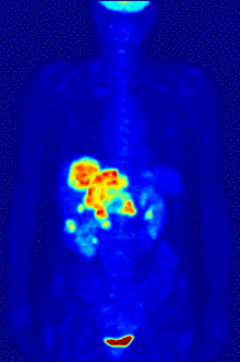
A radiopharmaceutical, such as FDG (fluorodeoxyglucose), that gives off gamma ray signals, is injected into the patient and its emissions are detected by a PET scanner. It measures the amount of metabolic activity at various sites in the body and a computer reassembles the signals into images.
Cancer cells have higher metabolic rates than normal cells, and show up as denser areas on a PET scan. It is useful in diagnosing certain cardiovascular and neurological diseases because it highlights areas with increased, diminished or no metabolic activity.
What preparations are needed for undergoing a PET-Scan?
For a PET-CT scan a patient needs to spare approximately 2-3 hrs. He/She should report to the PET CT facility essentially after prior appointment in a fasting state of approximately 6 hrs. In case of diabetics, the blood sugar levels should be controlled so that the fasting blood sugar is less than 175 mg/dL. The PET scan may be non-conclusive if performed under higher blood sugar levels.
How can a PET scan make a difference in cancer management?
PET being a metabolic imaging tool, can characterize a tumor as metabolically active or inactive, thereby obviating the need for surgical biopsy when the PET scan is negative. In case of active metabolism in a tumor, it helps in localizing the site of disease for accurate tissue sampling.
A PET scan facilitates the demonstration of abnormal metabolism in cancer cells when there is no anatomical abnormality detectable on CT. It is extremely useful in determining the full extent of a malignant disease. Lymph nodes when small in size can commonly be missed or tend to appear normal on a routine CT scan, aiding in the detection of lymphatic metastasis.
What are the other uses of a PET CT scan?
PET scan is considered as the gold standard to demonstrate metabolic activity in the myocardium. This is particularly useful in patients after myocardial infarction/heart attack before subjecting them to revascularization procedures. A dead (non-viable) myocardium is not expected to improve in function even after restoration of the blood circulation by angioplasty or a bypass surgery. On the contrary, if the infarcted myocardium has preserved metabolism (viable-hibernating) it shows dramatic improvement in function. PET scans are also useful in various neurological conditions, especially dementias and epilepsy where various areas of the brain can be screened for abnormal metabolism.